Bovine Viral Diarrhoea (BVD)
BVD is a complicated and economically very important disease. It is a viraldisease that infects cattle, sheep, deer and goats and is present on 80-90% of NZfarms. It is estimated approximately 60% of cattle have been exposed to it at some time.
How is it spread?
The virus is spread in all body fluids including saliva, tears, nasal discharge,semen, urine and faeces. Close contact with other animals is required fortransmission and the incubation period is about 1-3 weeks. The virus only lives a shorttime in the environment.
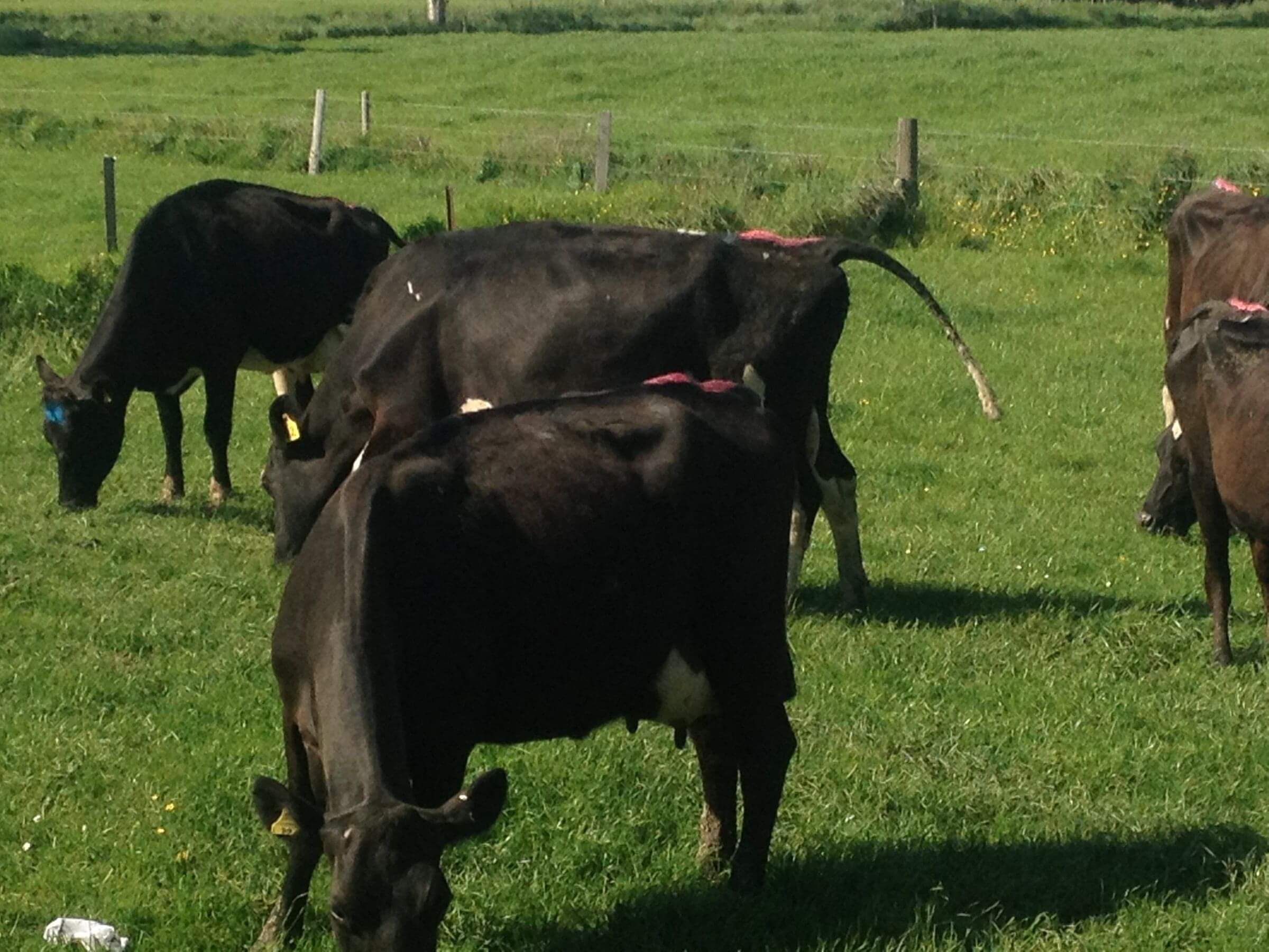
Signs of disease in a herd
In young cattle (3-12 months of age) symptoms include:
• scouring
• ulcers in the mouth with dribbling from the mouth
• ulcers between the toes causing lameness
• coughing
• nasal and eye discharge
• rough coat,
• reduced appetite with nil or poor weight gain and loss in body condition.
• some animals will be sub clinically affected and not show symptoms except
reduced weight gain.
BVD in young stock is frequently not diagnosed; because symptoms can be similar to parasitism. Farmers think their stock have worms and drench them without getting a diagnosis. As a lot of stock are recovering a month after getting infected, farmers get the false impression that their stock have responded to a drench when in fact they have not had worms at all.
In adult breeding cows the disease can cause huge economic reproductive wastage, the costs of which are often hidden and difficult to calculate.
Reproductive wastage occurs when a heifer or cow becomes exposed to the virus for the first time while she is pregnant. The effect seen depends on the stage of pregnancy the cow is infected at i.e.
- Day 10-30: Early embryo loss - seen as a reduced conception rate
- Day 30-125: The production of persistently infected (PI) animals happens here, these are born as apparently normal calves but shed the virus for life and can go onto to develop a fatal mucosal disease
- Day 100-150 May cause abnormal calves to be born e.g. hydrocephalus, cataracts, cleft palate, bent legs, still births etc
- From Day 180 Usually no effect - these calves are born with good immunity.
Diagnosis
Diagnosis
Diagnosis of PI animals can be made by blood or skin tests or by isolating the virus from the tissues of a dead animal. PI animals are virus (antigen) positive but antibody negative - i.e they are producing no immune response to the virus.
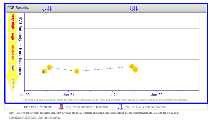
Bulk milk testing can be used to screen for the presence of historic and current levels of infection within a herd of dairy cows. Blood testing can be used to screen a mob of calves. Previously infected animals i.e animals that have been exposed to the virus after birth will be negative for the virus (antigen negative) but antibody positive. Antibodies remain present in herd test results for a long time after the infective PI animal has been removed.
Virus Elimination and Control
PI animals
These animals are infected for life and are the ones who spread the disease to the rest of the herd. Their contact with pregnant cows is what allows the next generation of PI calves to be born. Therefore they must be culled
Screening
Biosecurity and testing of any incoming stock is important . Bulls are often the big focus here but so too should be any in calf animal that is purchased whose herd history is uncertain. Brought in calf animals have the potential to already be carrying a PI calf inside them without you knowing.
Vaccination
Vaccination is an insurance policy against disease. In herds that have very low levels of antibody showing on bulk milk screening vaccination provides protection for naive animals against any incoming PI animals.
Vaccination of heifers moving to grazing where mobs of cattle from different farms could be mixed is advised